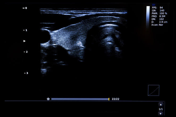
What is a Thyroid Ultrasound scan?

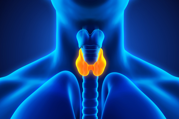
An ultrasound of the thyroid produces pictures of the thyroid gland and the adjacent structures in the neck. The thyroid gland is located in front of the neck just above the collar bones and is shaped like a butterfly, with one lobe on either side of the neck connected by a narrow band of tissue. It is one of nine endocrine glands located throughout the body that make and send hormones into the bloodstream.

The thyroid gland makes the thyroid hormone, which helps to regulate a variety of body functions including how fast the heart beats. It is very common for patchy areas or nodules to develop in the thyroid that may or may not be felt on the skin surface. About five to 10 percent of adults will have lumps in their thyroid that a doctor can identify on an exam. These are called palpable nodules. Ultrasound is very sensitive and shows many nodules that cannot be felt. In some age groups, nodules are seen on ultrasound in as many as 70 percent of adults. The vast majority of these are benign regions of thyroid tissue that pose no health risk. The minority of these are true tumors of the thyroid and may require further diagnosis or treatment.

A Thyroid Ultrasound may be used to:

  • determine if a lump in the neck is arising from the thyroid or an adjacent structure
  • to analyze the appearance of thyroid nodules and determine if they are the more common benign nodule or if the nodule has features that require a biopsy. If biopsy is required, ultrasound-guided fine needle aspiration can help improve accuracy of the biopsy.
  • to look for additional nodules in patients with one or more nodules felt on physical exam
  • to see if a thyroid nodule has substantially grown over time

For most ultrasound exams, you will be positioned lying face-up on an examination table that can be tilted or moved. Patients may be turned to either side or on occasion placed in a face down position to improve the quality of the images.

A pillow may be placed behind the shoulders to extend the area to be scanned for a thyroid ultrasound exam. This is especially important for a small child with very little space between the chin and the chest.

After you are positioned on the examination table, the radiologist or sonographer will apply a warm water-based gel to the area of the body being studied. The gel will help the transducer make secure contact with the body and eliminate air pockets between the transducer and the skin that can block the sound waves from passing into your body. The transducer is placed on the body and moved back and forth over the area of interest until the desired images are captured.

There is usually no discomfort from pressure as the transducer is pressed against the area being examined. However, if scanning is performed over an area of tenderness, you may feel pressure or minor pain from the transducer.

Once the imaging is complete, the clear ultrasound gel will be wiped off your skin. Any portions that are not wiped off will dry to a powder. The ultrasound gel does not stain or discolor clothing.

Your patient's exam will be interpreted by a board certified radiologist, who dictates a report. You will then receive your results within 48 to 72 hours.

Read about ultrasound for the following specialties:

Ophthalmology

Read more now

Cardiac & Vascular

Read more now

Internal Medicine

Read more now

Schedule a Free Consultation with ProVision today

and find out how we can help to increase your bottom line!

SCHEDULE NOW