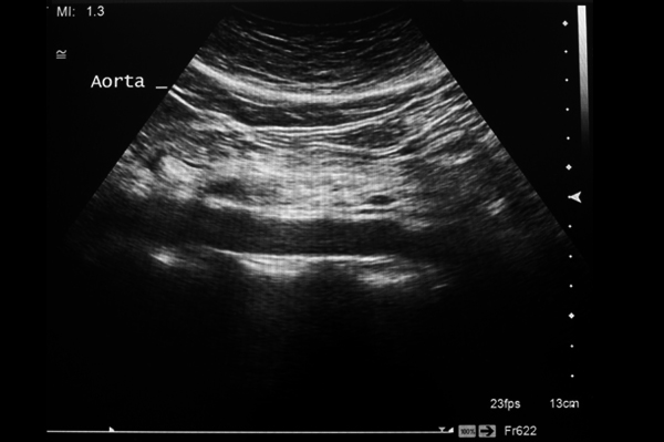
According to the American Vascular Association, more than 17,000 Americans die of ruptured abdominal aortic aneurysms each year. Abdominal Aortic Aneurysms are such a high health risk because people have little or no symptoms. The AAA ultrasound will quickly and accurately identify any abnormalities.
A duplex ultrasound can help diagnose the following conditions:
- Abdominal aneurysm
- Arterial occlusion
- Blood clot
- Carotid occlusive disease (See: Carotid duplex)
- Renal vascular disease
- Varicose veins
- Venous insufficiency
Some preparation is needed. The study examines arteries deep in the abdomen. Gas in the intestinal tract can interfere with ultrasound evaluation. It is therefore best to have the examination performed after an overnight fast, and it is important to avoid tobacco and caffeine prior to the test. A complete study can take an hour or two. Scanning may be performed from the front or sides of the abdomen and can be facilitated by the patient lying on one side or the other.
Evaluation by a vascular surgeon will generally be recommended if there is a renal artery narrowing of 60 percent or more. Further evaluation or treatment may be recommended. Intervention may be appropriate if renal artery narrowing is thought to be contributing to blood-pressure problems, or if severe narrowing threatens the continued function of the kidney. Renal artery stenting is the most common intervention offered when treatment is needed, but some patients may need a surgical procedure to address complex renal artery disease.
For patients found to have only mild to moderate renal artery disease, a follow-up study in the Vascular Laboratory offers a safe, non-invasive and accurate means to assess for progression of renal artery disease over time.

Your patient's exam will be interpreted by a board certified radiologist, who dictates a report. You will then receive your results within 48 to 72 hours.


