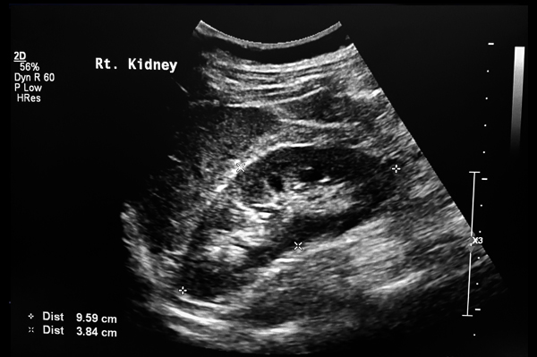
A kidney ultrasound may be used to:
- Assess the size, location, and shape of the kidneys and related structures, such as the ureters and bladder.
- Detect cysts, tumors, abscesses, obstructions, fluid collection, and infection within or around the kidneys.
- Detect calculi (stones) of the kidneys and ureter.
- Assist in placement of needles used to biopsy (obtain a tissue sample) the kidneys and drain a cyst.
- To evaluate a transplanted kidney.
- See how blood is flowing to the kidneys through the renal arteries and veins.
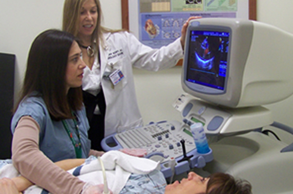
A kidney ultrasound may be performed on an outpatient basis or as part of your stay in a hospital. You will be asked to remove any clothing, jewelry, or other objects that may interfere with the scan. If asked to remove clothing, you will be given a gown to wear.
You will lie on an examination table on your stomach. Ultrasound gel is placed on the area of the body that will undergo the ultrasound examination. Using a transducer, a device that sends out the ultrasound waves, the ultrasound wave will be sent through that your body. The sound will be reflected off structures inside the body, and the ultrasound machine will analyze the information from the sound waves. The ultrasound machine will create images of these structures on a monitor. These images will be stored digitally.
If the bladder is examined, you will be asked to empty your bladder after scans of the full bladder have been completed. Additional scans will be made of the empty bladder.
There are no confirmed adverse biological effects on patients or instrument operators caused by exposures to ultrasound at the intensity levels used in diagnostic ultrasound.

Your patient's exam will be interpreted by a board certified radiologist, who dictates a report. You will then receive your results within 48 to 72 hours.