You may need this scan if your health care provider thinks you may have:
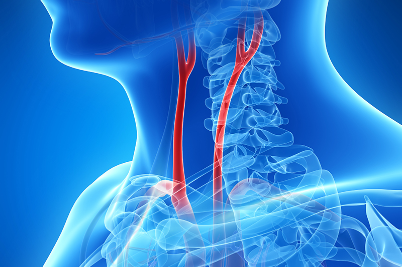
- A blockage (occlusion) in a carotid artery
- Narrowing (stenosis) in a carotid artery
- A blockage may be caused by a buildup of fatty material (plaque), a blood clot (thrombus), or other substances.
Symptoms of blockage may include:
- Dizziness
- Confusion
- Drowsiness
- Headache
- Temporary blindness in one eye
- Temporary inability to speak or move
- These symptoms may be early warning signs of a stroke.
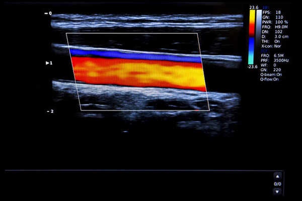
You may also need this scan even if you have no symptoms but your health care provider hears an abnormal sound (bruit) in an artery. This abnormal sound may mean that you have a problem with blood flow in the artery.
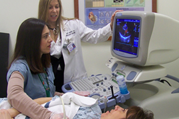
Some preparation is needed. The study examines arteries deep in the abdomen. Gas in the intestinal tract can interfere with ultrasound evaluation. It is therefore best to have the examination performed after an overnight fast, and it is important to avoid tobacco and caffeine prior to the test. A complete study can take an hour or two. Scanning may be performed from the front or sides of the abdomen and can be facilitated by the patient lying on one side or the other.
Evaluation by a vascular surgeon will generally be recommended if there is a renal artery narrowing of 60 percent or more. Further evaluation or treatment may be recommended. Intervention may be appropriate if renal artery narrowing is thought to be contributing to blood-pressure problems, or if severe narrowing threatens the continued function of the kidney. Renal artery stenting is the most common intervention offered when treatment is needed, but some patients may need a surgical procedure to address complex renal artery disease.
For patients found to have only mild to moderate renal artery disease, a follow-up study in the Vascular Laboratory offers a safe, non-invasive and accurate means to assess for progression of renal artery disease over time.
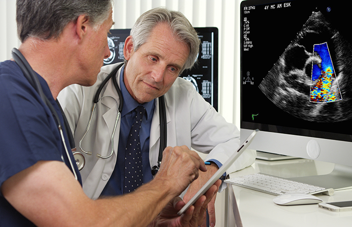
Your patient's exam will be interpreted by a board certified radiologist, who dictates a report. You will then receive your results within 48 to 72 hours.