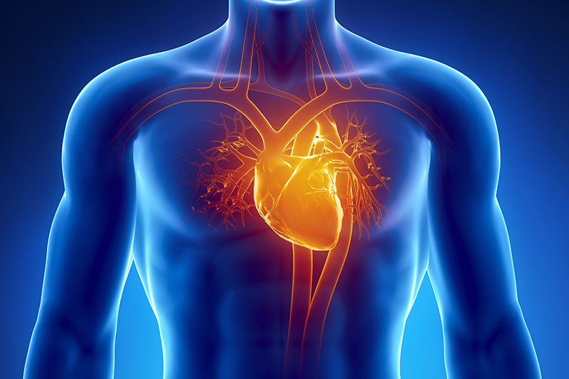
- Assess the overall function of your heart.
- Determine the presence of many types of heart disease.
- Follow the progress of heart valve disease over time.
- Evaluate the effectiveness of medical or surgical treatments.
- Valvular heart disease - valve dysfunction, follow-up of prosthetic valves.
- Abnormal left ventricular function - used to assess any underlying cause and to estimate left ventricular ejection fraction (LVEF).
- Atrial fibrillation- assess structural cause, risk of thromboembolism and likely response to direct current (DC) cardioversion.
- Congenital heart disease.
- Cardiomyopathy.
- Infective endocarditis - including assessment of valvular lesions and their haemodynamic severity.
- After embolic stroke - assess possible cardiac source.
Pericardial disease - presence of fluid and allows guided (and therefore safe) drainage of pericardial fluid in cardiac tamponade.[2]
Thoracic aortic disease - aneurysm, dissection (although CT is an alternative).
An Echocardiogram usually takes less than an hour to do. For some types of echo, your doctor will need to inject saline or a special dye into one of your veins. The substance makes your heart show up more clearly on the echo pictures. The dye used for echo is different from the dye used during angiography (a test used to examine the body's blood vessels).
For most types of echo, you will remove your clothing from the waist up. Women will be given a gown to wear during the test. You'll lie on your back or left side on an exam table or stretcher. Soft, sticky patches called electrodes will be attached to your chest to allow an EKG (electrocardiogram) to be done. An EKG is a test that records the heart's electrical activity.
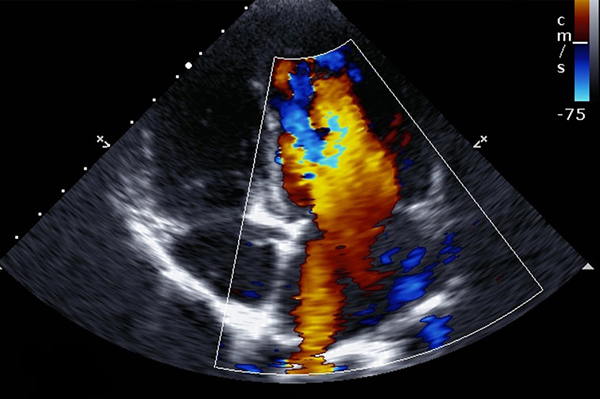
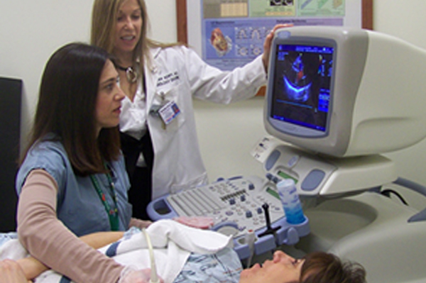
A doctor or sonographer (a person specially trained to do ultrasounds) will apply gel to your chest. The gel helps the sound waves reach your heart. A wand-like device called a transducer will then be moved around on your chest. The transducer transmits ultrasound waves into your chest. A computer will convert echoes from the sound waves into pictures of your heart on a screen. During the test, the lights in the room will be dimmed so the computer screen is easier to see.

Your patient's exam will be interpreted by a board certified radiologist, who dictates a report. You will then receive your results within 48 to 72 hours.