- Find the cause of abdominal pain
- Find the cause of kidney infections
- Diagnose a hernia
- Diagnose and monitor tumors and cancers
- Diagnose or treat ascites
- Learn why there is swelling of an abdominal organ
- Look for damage after an injury
- Look for stones in the gallbladder or kidney
- Look for the cause of abnormal blood tests such as liver function tests or kidney tests
Look for the cause of a fever
For an abdominal ultrasound exam, you will be positioned lying face-up on an examination table that can be tilted or moved. Patients may be turned to either side or on occasion placed in a face down position to improve the quality of the images.
After you are positioned on the examination table, the radiologist or sonographer will apply a warm water-based gel to your abdominal area. The gel will help the transducer make secure contact with the body and eliminate air pockets between the transducer and the skin that can block the sound waves from passing into your body. The transducer is placed on the body and moved back and forth over the area of interest until the desired images are captured.
There is usually no discomfort from pressure as the transducer is pressed against the area being examined. However, if scanning is performed over an area of tenderness, you may feel pressure or minor pain from the transducer.
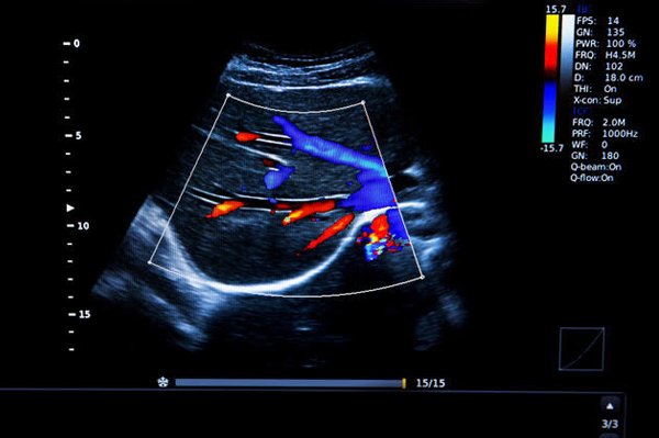
Doppler sonography is performed using the same transducer.
Once the imaging is complete, the clear ultrasound gel will be wiped off your skin. Any portions that are not wiped off will dry to a powder. The ultrasound gel does not stain or discolor clothing.

Your patient's exam will be interpreted by a board certified radiologist, who dictates a report. You will then receive your results within 48 to 72 hours.